“A.I. could play a big role in supporting prevention, diagnosis, treatment plans, medication management, precision medicine and drug creation” __Bruce Liang, Chief Information Officer of Singapore’s Ministry of Health
In software development, versioning is one of the key tenets of good programming. One can go back in history using a version control system such as git, svn, cvs etc to troubleshoot bugs, reverse deployment. Wouldn’t it be cool if a similar system existed in medical imaging which can assist radiologists to quickly “see” if a treatment is positively or negatively affecting the patient? Computer vision can process images and highlight differences between two or more images in real-time. That means, a radiologist need not spend hours retrieving, interpreting images of a patient and identifying the differences, with a click of a button on their phone they can see highlights of what has changed between images.
If there was a hypothetical medical imaging versioning system, how would such a system work, how would it be implemented and deployed in hospital systems, who would primarily use it, how would it enhance treatment effectiveness?
“Medical imaging guides the course of much of patient care and is an essential element of biomedical research. From x-rays and ultrasound to computerized tomography (CT), functional magnetic resonance imaging (fMRI), and positron emission tomography (PET), medical imaging helps clinicians diagnose, treat, and understand a range of diseases and conditions, including cancer, cardiovascular disease, and neurodegenerative disorders.”
Roadmap for Medical Imaging Whitehouse.gov
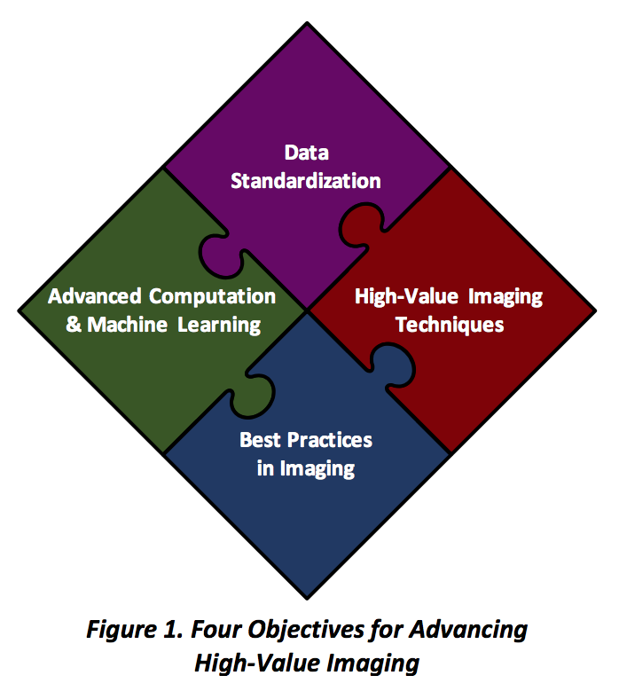
Internet Working Group for Medical Imaging (IWDMI) defined the above as the four key pieces for better healthcare through effective medical imaging. I’m particularly interested in “Advanced Computation & Machine Learning” aspect of the roadmap.
Here is a set of breast cancer images for a patient taken at regular intervals. I’m not going to pretend to know what’s going on in the following image but anyone with half a brain can guess that it shows images of different stages of breast cancer and it’s trying to help the physician understand treatment’s effectiveness over time (week 1 through week 20).
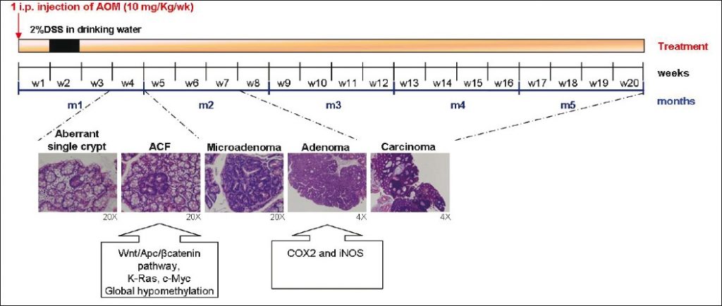
For a radiologist to pull such a report, my sense is, it’s not straightforward. Retrieving images from disparate systems, putting them next to each other for quick and easy comparison and looking at the treatments (dosage etc) alongside the images and viewing that over time to get a sense of disease progression probably takes hours if not days.
This can be streamlined and automated using better image storage, retrieval and computer vision. If we can reduce the amount of time to generate this report from days/hours to minutes/seconds, it would save precious time for physicians and might be a life saver for the patient.
In this series, next we will see where the current art is on this issue and subsequently look at the possibilities of using latest Computer Vision (CV) techniques to save time for Radiologists and Pathologists.